Boletus L.
Recent molecular studies have shown that Boletus in its current circumscription is likely an artificial grouping and it is possible that it will be split at some point into smaller genera. Note that Boletus impolitus and Boletus depilatus for practical reasons are retained here, although there is strong evidence that they are closely related to Xerocomus subtomentosus and its allies.
Fruitbody large to medium sized, boletoid, without veil and ring. Stipe solid, with surface usually covered with granules or network. Flesh variously coloured, changing or not when exposed to air. Tubes easily separable from each other, not tearing apart. Pores usually small and rounded.
Boletus adalgisae Marsico & Musumeci
Description
Pileus up to 8 cm, convex to flat-convex, flat or depressed, velvety to smooth, reddish brown, pinkish brown or brown, in places discolouring, in old fruitbodies with purple tinge, blueing when bruised. Stipe cylindrical, tapering towards the base or sometimes slightly ventricose or nearly clavate, pale yellow to yellow, covered with fine reddish granules, in the base vinaceous brown; stipe surface blueing when bruised. Flesh whitish, pale yellow to lemon yellow, vinaceous red to vinaceous brown in the stipe base, blueing when exposed to air. Tubes pale yellow to yellow with olivaceous tint, blueing when injured. Pores red to orange red or orange, blueing when bruised. Smell and taste not recorded. Spores 12–17.5 × 6.5–9 μm, mean ratio ca 1.9, amyloid. Pileipellis (the cap cuticle) a trichoderm, composed of hyphae of cylindrical cells. Chemical reactions: hyphae of the flesh in the stipe base inamyloid in Melzer’s reagent.
Habitat. Warm broadleaf forests, mycorrhizal with oaks (Quercus), sweet chestnut (Castanea sativa) or beech (Fagus).
Distribution. Described and so far known only from Italy.
Similarity. Apparently similar to Boletus queletii, but distinguished by the non-amyloid hyphae of the flesh in the stipe base, amyloid spores (unique feature among the European members of the genus) and the peculiar cystidia coloured ochraceous in KOH and Melzer’s solution.
Photographs
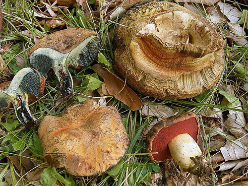
Boletus adalgisae - basidiomata in different stages of development. Photo O. Marsico.
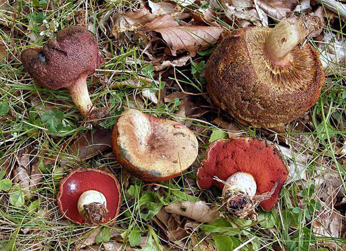
Basidiomata of Boletus adalgisae. Note the tubes. (phoro O. Marsico)
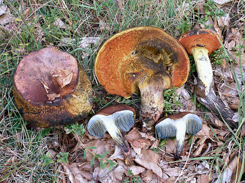
Fruitbodies of Boletus adalgisae. Note the superficial resemblance to Boletus queletii. (photo O. Marsico)
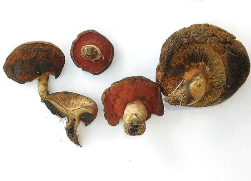
Fruitbodies of Boletus adalgisae. (photo O. Marsico)
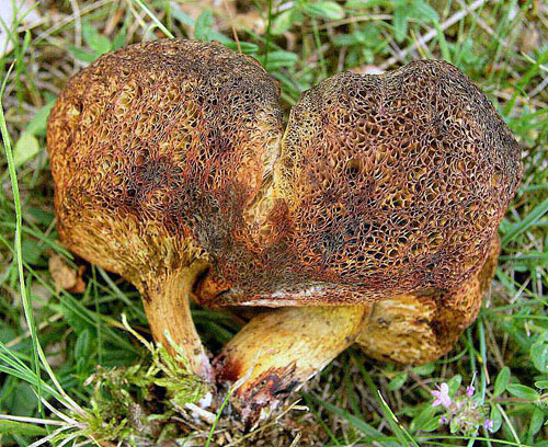
Mature fruitbodies of Boletus adalgisae - view of the stipe and the tubes. (photo O. Marsico)
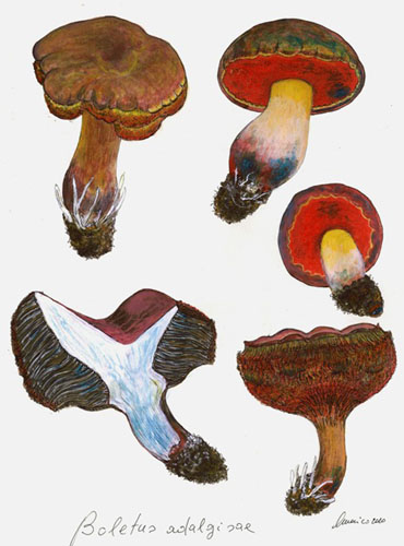
Fruitbodies of Boletus adalgisae. (painting O. Marsico)
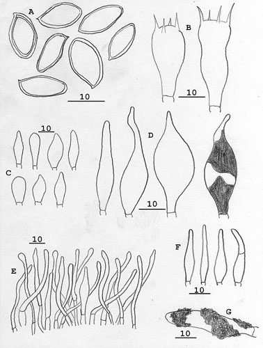
Boletus adalgisae - microscopic features: a - spores, b - basidia, c - cells from the tubes margin, d - pleurocystidia, e - pileipellis, f, g - caulocystidia. Scalebars = 10 μm. (drawing E. Musumeci)
Important literature
Marsico, O. & Musumeci, E. 2011. Boletus adalgisae sp. nov. – Bolletino dell’Associazione Micologica ed Ecologica Romana 27: 3–15.