Xerocomus Quél.
Recent molecular studies have shown that Xerocomus in its current circumscription is likely an artificial grouping and it is possible that it will be split at some point into smaller genera. Molecular studies also have changed our understanding about the species of xerocomoid boletes showing that morphological features are quite variable in this group. Not only microscopic study is essential for determination, but scanning electron microscope will be often needed in this “genus” as the spore ornamentation is not always seen under ordinary light microscope. Do bear in mind that macroscopic characters, such as colours, cracking cuticle, etc., tend to intergrade between the different species. Note that Boletus impolitus and Boletus depilatus that were shown to be close to Xerocomus subtomentosus and its allies, are here retained in Boletus for practical reasons. The same applies also for Phylloporus pelletieri, placed here in a genus of its own, but being also close to Xerocomus subtomentosus group.
Although large reference list will be found under most of the species, one should always consult Ladurner & Simonini (2003) having in mind that there are some new species (X. chrysonemus, X. marekii, X. silwoodensis) described after this otherwise superior book was printed. Useful keys, covering most of the European xerocomoid boletes (except some southern taxa) are provided by Knudsen & Vesterholt (2008), Hills (2008) and Kibby (2011), the later also featuring an excellent comparison chart.
Fruitbody medium to small sized, boletoid, without veil and ring. Stipe solid, often tapering towards the base. Flesh variously coloured, changing or not when exposed to air. Tubes not separable from each other, instead tearing apart. Pores usually angular.
Xerocomus chrysonemus A.E. Hills & A.F.S. Taylor
Description
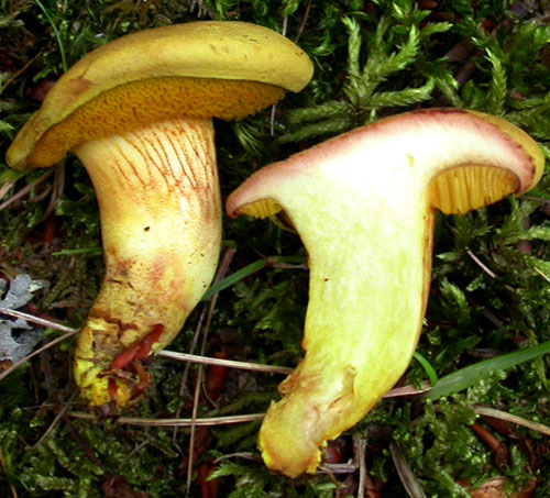
This recently described species, associated with oaks (Quercus), is known to me only from the descriptions found in the literature. Morphologically it is very similar to Xerocomus subtomentosus and X. ferrugineus. From X. subtomentosus it is distinguished by the bright yellow (not pinkish or brownish) flesh in the stipe base that is usually not changing when exposed to air (instead of the flesh of X. subtomentosus that is normally blueing in the cap. Also the basal mycelium is yellow to bright yellow (whitish in X. subtomentosus). Xerocomus ferrugineus also has yellowish mycelium but it has whitish unchanging flesh and often grows under conifers. There are some differences also in the sizes of the spores, explained in detail in the paper with the original description of X. chrysonemus (Taylor & al. 2006; available online).
Habitat. In damp shady places, mycorrhizal with oaks (Quercus).
Distribution. Not yet understood, so far known only from the United Kingdom and Spain. Should be further looked for.
Photographs

Fruitbody of Xerocomus chrysonemus. The yellow flesh in the stipe base is important characteristic of this species. (photo A. E. Hills)
An illustration of the microscopic features is found in the paper of Taylor & al. (2006; click here to read online). Spanish collections with SEM microphotographs are illustrated in Muñoz et al. (2008; available online).
Important literature
Gelardi, M. 2011. A noteworthy British collection of Xerocomus silwoodensis and a comparative overview on the European species of X. subtomentosus complex. – Bolletino dell’ Associazione Micologica ed Ecologica Romana 84: 28–38.
Hills, A.E. 2008. The genus Xerocomus. A personal view, with a key to the British species. Field Mycology 9(3): 77–96.
Knudsen, H. & Vesterholt, J. [eds.]. 2008. Funga Nordica. Nordsvamp, Kopenhagen.
Muñoz, J.A., Cadiñanos Aguirre, J. A. & Fidalgo, E. 2008. Contribución al catálogo corológico del género Xerocomus en la Peninsula Iberica. – Boletín de la Sociedad Micológica de Madrid 32: 249–277. (available online)
Taylor, A.F.S., Hills, A.E., Simonini, G., Both, E.E. & Eberhardt, U. 2006. Detection of species within the Xerocomus subtomentosus complex in Europe using rDNA–ITS sequences. – Mycological research 110: 276–287.